WHAT IS APDS?

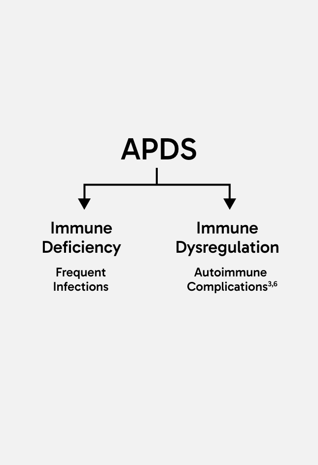
Activated PI3Kδ syndrome (APDS) is a rare type of inborn error of immunity (IEI), which is characterised by both immune deficiency and immune dysregulation.1-3

First described in 2013, APDS was previously known as PASLI* disease and is one of a group of diseases sometimes known as primary immunodeficiencies (PIDs).1-6

Clinical manifestations of APDS typically begin in childhood and get progressively worse over time, often leading to irreversible organ damage, lymphoma and early mortality.2-4,7

four cartoon people

Research suggests that approximately one in four people with APDS face early mortality by the age of 30 years old.8

40%

of patients have a family history of IEIs.

In almost 40% of cases, patients have a family history of IEIs,9 however APDS can present spontaneously with neither parent being a carrier of the genetic variants which cause the disease.10 It is estimated that APDS affects one to two people per million, including both adults and children.11
GENETIC BASIS OF APDS

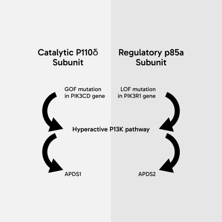
APDS is caused by variants in either one of two genes encoding phosphoinositide 3-kinase delta (PI3Kδ), which are vital for the development and function of immune cells.1

  • Variants of PIK3CD lead to gain of function in the P110δ catalytic subunit, resulting in APDS11
  • Variants of PIK3R1 lead to loss of function in the p85α regulatory subunit, resulting in APDS21,12
Diagram showing that a gain of function mutation in the PIK3CD gene, which encodes the catalytic P110δ subunit, leads to hyperactivation of the P13K pathway, resulting in APDS1. Diagram also shows that a loss of function mutation in the PIK3R1 gene, which codes for the regulatory p85a subunit, leads to hyperactivation of the P13K pathway, resulting in APDS2.

The disturbance of homeostatic PI3K signalling leads to abnormalities in immune cell development, cellular metabolism, maturation and proliferation, resulting in both immune deficiency and immune dysregulation.13

Clinical manifestations of APDS

Over time, immune deficiency leads to frequent or chronic respiratory / sinopulmonary infections, which can lead to bronchiectasis. It also predisposes to herpes virus infections such as Epstein-Barr virus (EBV) and cytomegalovirus (CMV).3,6

Prolonged immune dysregulation can lead to lymphoproliferation, including lymphadenopathy and splenomegaly, enteropathy, autoimmune disorders and lymphoma.3,6

Patients with APDS typically experience a variety of acute and chronic clinical features within multiple organ systems.3,9,10,14 However, not all patients with APDS experience the same clinical profile, even those within the same family.11

Simple line hierarchy diagram with the term APDS at the top and the line splits to show it leads to both immune deficiency which leads to frequent infections and immune dysregulation which leads to autoimmune complications.

Diagnosing APDS

The broad spectrum of clinical manifestations of APDS affecting multiple organ systems over a prolonged period can result in a delayed diagnosis.9

In order to prevent long term complications and early mortality resulting from APDS, timely and accurate diagnosis is key.7

Three cartoon people

The only definitive way to diagnose APDS is via a genetic test.15

7YRS TO DIAGNOSIS

On average, it takes seven years for a patient to be diagnosed with APDS.9

*P110d-Activating mutation causing Senescent T cells, Lymphadenopathy and Immunodeficiency.

  1. Lougaris V, et al. Pediatr Allergy Immunol. 2022;33 Suppl 27:69-72.
  2. Elkaim E, et al. J Allergy Clin Immunol. 2016;138(1):210-218.e9.
  3. Coulter TI, et al. J Allergy Clin Immunol. 2017;139(2):597-606.e4.
  4. Angulo I, et al. Science. 2013;342(6160):866-871.
  5. Rao KV, et al. Blood. 2017:130(21):2307-2316.
  6. Rao KV, et al. Blood. 2023;141(9):971-983.
  7. Redenbaugh V, Coulter T. Front Pediatr. 2021;9:702872.
  8. Hanson J, Bonnen PE. J Clin Exp Med. 2024;24:17.
  9. Jamee M, et al. Clin Rev Allergy Immunol. 2020;59(3):323-333.
  10. Deau MC, et al. [published correction appears in J Clin Invest. 2015 Apr;125(4):1764-5]. J Clin Invest. 2014;124(9):3923 -3928.
  11. Vanselow S, et al. Front Immunol. 2023 ;14. :1208567.
  12. Lucas CL, et al. J Exp Med. 2014;211(13):2537-47.
  13. Cant AJ, et al. J Allergy Clin Immunol Pract. 2024;12(1):69-78.
  14. Coulter TI, Cant AJ. Front Immunol. 2018;9:2043.
  15. Chinn IK, et al. J Allergy Clin Immunol. 2020;145(1):46-69.