DIAGNOSING APDS

APDS is difficult to diagnose due to its varied clinical manifestations within multiple organ systems.1

The only definitive way to diagnose APDS is via a genetic test.2

APDS can be inherited in an autosomal dominant pattern, meaning that a person can inherit a variant gene from only one parent for the disease to manifest.3 Children born to a parent with variants in either one of two genes encoding phosphoinositide 3-kinase delta (PI3Kδ) will have a 50% chance of inheriting APDS.4

Simple human figure infographics in different sizes – small, medium and large. The small and medium figures are coloured black, and the large figure is coloured white.
50% CHANCE AN AFFECTED PARENT WILL PASS AN APDS GENE ON TO THEIR CHILD.

Family members of patients with a confirmed IEI/APDS diagnosis should be recommended for genetic testing with a panel that includes APDS.

A waist-up shot of a male child and his dad washing and drying mugs in the sink together at home.

While almost 40% of patients have a family history of IEIs,4 APDS can also present spontaneously with neither parent being a carrier of the genetic variants which cause the disease.5

Not all patients with APDS look the same – signs and symptoms can vary, even among affected family members.6

Signs and symptoms of APDS typically begin in childhood and get progressively worse over time.1,4

Patients typically experience a variety of acute and chronic clinical features within multiple organ systems.4,7 It is important to look out for the wide range of clinical manifestations associated with APDS, as the onset of signs and symptoms can be relative to a patient’s age.

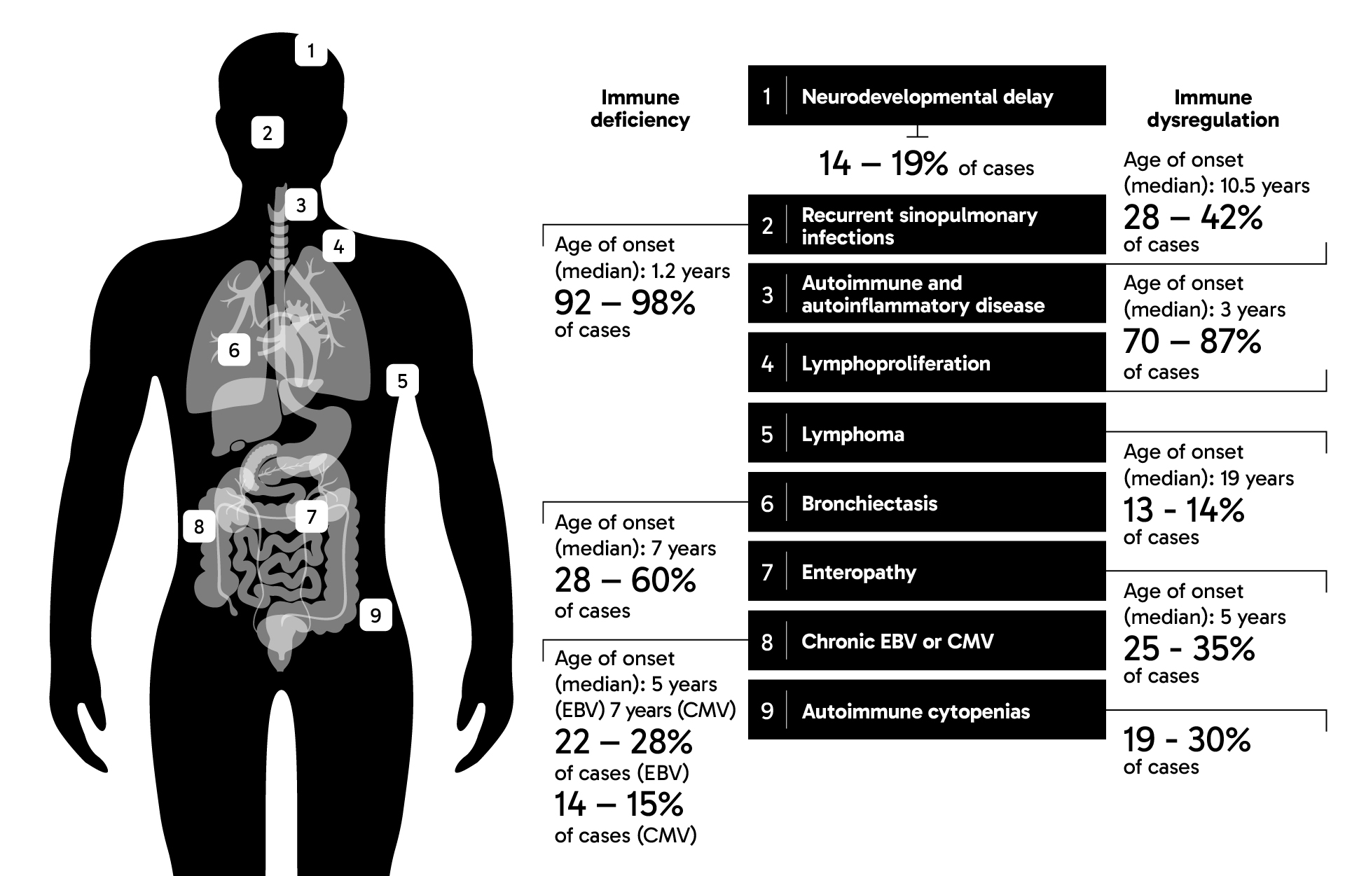
Clinical features of APDS typically include:1,4,8,9

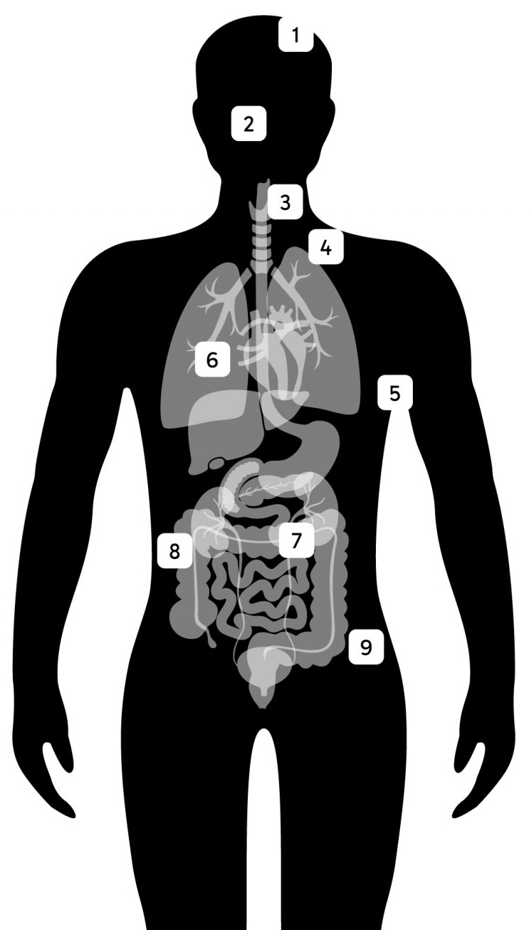
Features of APDS graphic

People with APDS usually experience the following clinical features (n≤243):1,4,7,8,10

Black and white infographic with a human figure with numbered pointers highlighting different body parts typically affected by APDS on the left and a list of APDS clinical features are shown on the right along with percentage of cases and median age of onset of each feature: The head of the figure is pinpointed 1 to show that neurodevelopment delay occurs in 14-19% of APDS cases. A number of clinical features of APDS are listed as resulting from immune deficiency: • The figure’s face is pinpointed 2 to show that severe, recurrent sinopulmonary infections occur in 92-98% of APDS cases, with a median age of onset of 1.2 years • The lungs are pinpointed 6 to show that bronchiectasis occurs in 28-60% of cases, with a mean age of onset of 7 years • The abdomen is pinpointed 8 to show that chronic EBV infections occur in 22-28% of APDS cases, with a median onset of 5 years, and chronic CMV infections occur in 14-15% of cases, with a median onset of 7 years Other APDS manifestations are listed as resulting from immune dysregulation, including: • The throat is pinpointed 3 to show that autoimmune and autoinflammatory disease occurs in 28-42% of cases, with a median age of onset of 10.5 years • The upper chest is pinpointed 4 to show that lymphoproliferation occurs in 70-87% of cases, with a median onset of 3 years • The armpit is pinpointed 5 to show that lymphoma occurs in 13-14% of cases, with a median age of onset of 19 years • The intestines are pinpointed 7 to show that enteropathy occurs in 25-35% of cases, with a median age of onset of 5 years • The lower abdomen is pinpointed 9 to show that autoimmune cytopenias occur in 19-30% of cases

Due to diagnostic challenges, APDS frequently follows a pattern of multiple referrals, with some patients seeing several different specialists over an average of seven years, before a diagnosis is confirmed.4

Laboratory tests for people with suspected APDS or PIDs:1,3,4,7,10

Laboratory test Typical observation in suspected patients

Complete blood count with differential

Depending on the individual presentation, several values may or may not be outside the normal range, for example:
• Red blood cells will not be low unless the patient has cytopenias
• White blood cells levels may vary based on the presence of infection, though lymphopenia is common

Immunoglobulin levels

• Low to normal concentrations of IgG and IgA
• Normal or elevated concentration of IgM

Vaccine challenge

• Many patients have a reduced response

Flow cytometry

T and B cell phenotypes are often altered. Distinguishing findings may include:
• Increased transitional B cells (CD19+, CD38+)
• Reduced naive B cells
• Increased TFH cells (CD4+, CXCR5+, PD1+)
• Reduced naive T cells (CD4+ CD8+)
• Reversed CD4/CD8 ratio
• Reduced CD4+ T cells

Early diagnosis of APDS is crucial to help prevent disease progression, organ damage and early mortality.6

  1. Coulter TI, et al. J Allergy Clin Immunol. 2017;139(2):597-606.e4.
  2. Chinn IK, et al. J Allergy Clin Immunol. 2020;145(1):46-69.
  3. Lucas CL, et al. Nat Immunol. 2014;15(1):88-97. doi:10.1038/ni.2771.
  4. Jamee M, et al. Clin Rev Allergy Immunol. 2020;59(3):323-333.
  5. Deau MC, et al. [published correction appears in J Clin Invest. 2015 Apr;125(4):1764-5]. J Clin Invest.
  6. Vanselow S, et al. Front Immunol. 2023;14:1208567.
  7. Elkaim E, et al. J Allergy Clin Immunol. 2016;138(1):210-218.e9.
  8. Maccari ME, et al. Front Immunol. 2018;9:543.
  9. Maccari ME, et al. J Allergy Clin Immunol. 2023;152(4):984–96.
  10. Angulo I, et al. Science. 2013;342(6160):866-871.